Ultrasound Tomography Breast Scanner
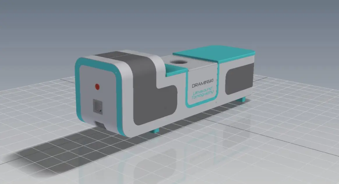
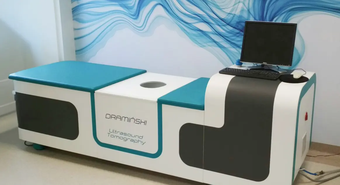
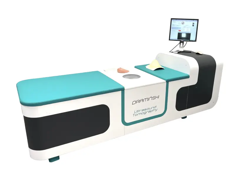
Ultrasound tomography as a non-invasive and safe hybrid method may contribute to achieving a new standard in breast cancer diagnostics. One of the few advanced prototypes of hybrid ultrasound tomography breast scanner in vivo being developed in the world was designed in Poland by DRAMIŃSKI S.A.
Read more
The future in 3D breast imaging
The device uses the ultrasound transmission tomography (UTT) method for quantitative imaging of female breast tissue structure, as well as a method of omnidirectional compounding multiple qualitative B-mode ultrasound images (FASCI – full angle spatial compound imaging).
Quick & Inexpensive
The scanner is designed for quick and inexpensive breast screening that will result in information about the presence, type and location of breast cancer lesions.
Safe & non-invasive
It is non-invasive and safe method that can contribute to achieving new standard in breast cancer diagnostics.
Differentiation
Ability to differentiate between benign and malignant lesions.
Radiation free
Breast examination is performed by scanning coronal slices every 1 mm along the breast lenght.
Introduction & Scanner Construction
- The device uses a computer algorithm to recognize breast cancer and estimate its malignancy to assist the diagnostic physician. There constructed quantitative distribution of the ultrasound velocity c [m/s] (UTTc) and the attenuation α [dB/cm] (UTTα) in the tissue structure of the breast volume scanned is used for the calculations.
- The scanner is designed for quick and inexpensive breasts screening that will result in information about the presence, type, and location of breast cancer lesions.
Scanner Functionality
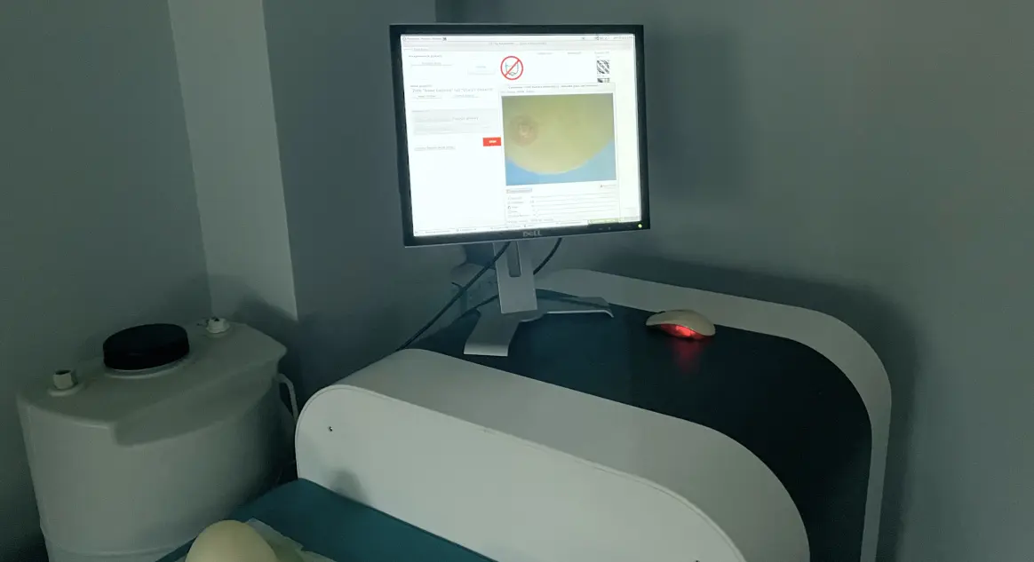
- Depending on the length of the breast, about 100 - 200 coronal cross-sections (slices) from the base to the nipple are examined. Scanning one cross-section currently takes about 6 s. Examination of the whole breast takes several minutes. It is still possible to reduce it by about three times. 3D visualization of structure of the breast tissue (in 3 different ultrasound modalities) is possible in several seconds just after scanning along with e.g. multi planar reconstruction (MPR) of 2D images in orthogonal sections (coronal, sagittal, and transverse).
Examination
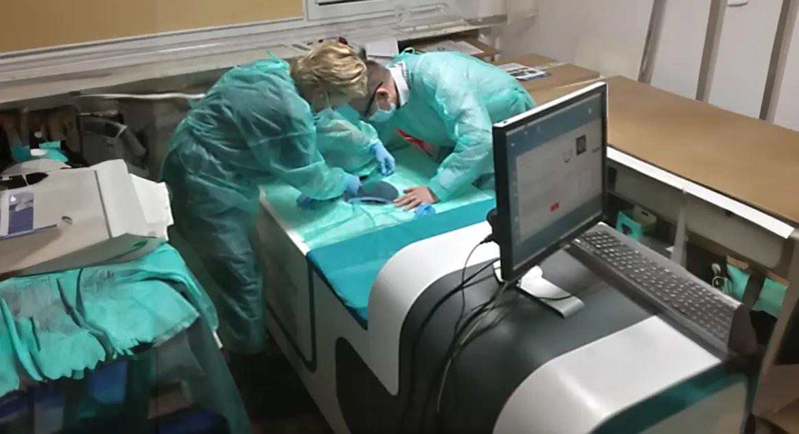
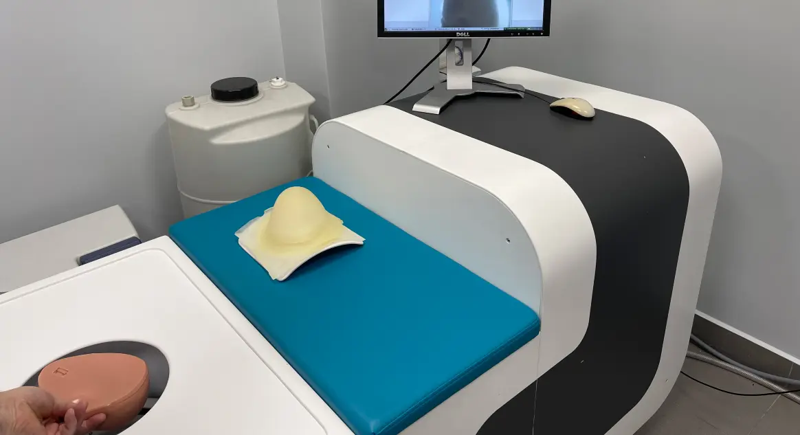
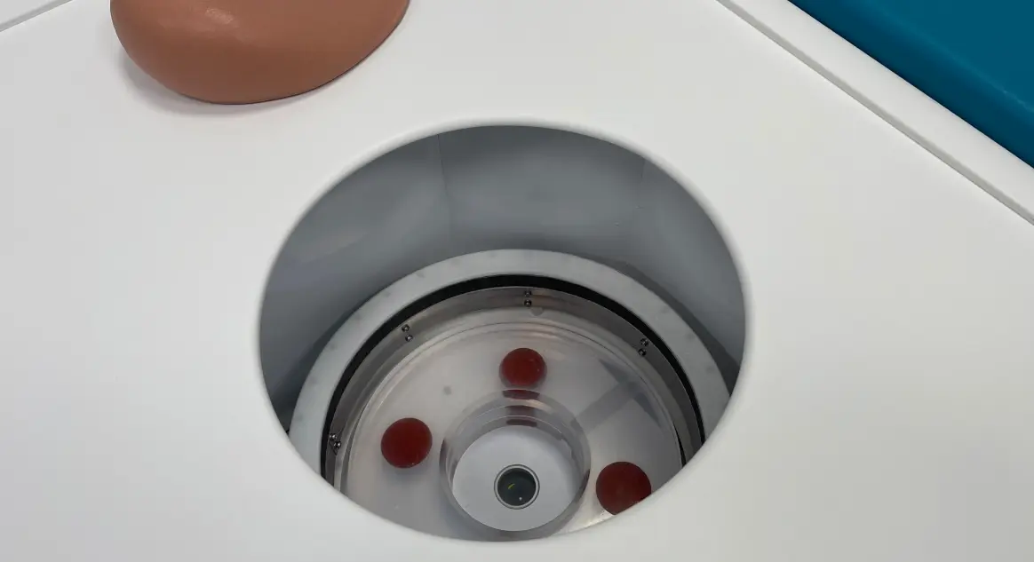
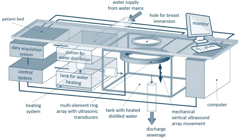
- The patient lies on her abdomen on the scanner bed with her breast submerged through the opening into a tank of distilled water, heated to approximately 25 °C. Breast examination is performed automatically by scanning coronal cross-sections (slices) every 1 mm or 2 mm, using a mechanically moving multi-element ring array in vertical direction, along the breast length.
Ultrasonic ring array
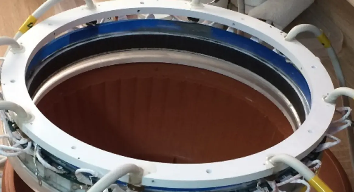
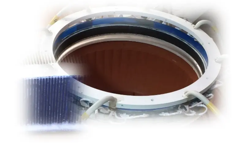
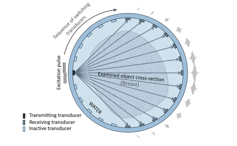
- The "heart" of the ultrasound tomography scanner is the ultrasonic ring array. It is made up of 1024 miniature piezo ceramic transducers evenly distributed on the inside of a 260 mm diameter ring, surrounding the examined breast. The elementary transducers have active area dimensions of 0.5 x 18 mm and operate with pulsed wave at approximately 2 MHz frequency. Each transducer can act as a transmitting and receiving element.
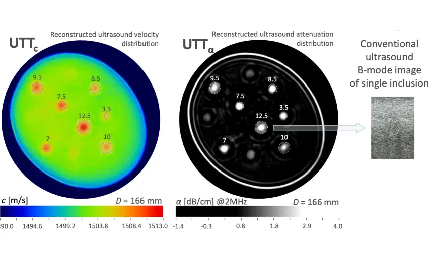
- Phantom inclusions have an acoustic impedance similar to the value in the surrounding gel and are invisible in the conventional ultrasound B-mode image. Similarly, they are very poorly see non the X-ray computed tomography image but highly visible on ultrasound transmission tomography images (UTTc and UTTα).
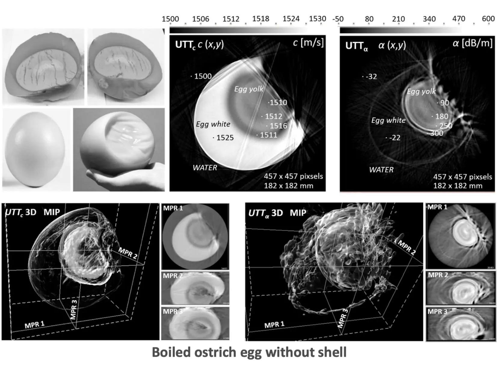
- The ultrasound velocity and attenuation values obtained are as expected: the yolk layers are clearly recognizable, whereas only the central region of the yolk ball with a lighter hue is visible in the optical images. The line artifacts on the UTT images are mainly due to the disappearance of the transmitted pulses by the disruption of the egg surface smoothness.
Ultrasound transmission images
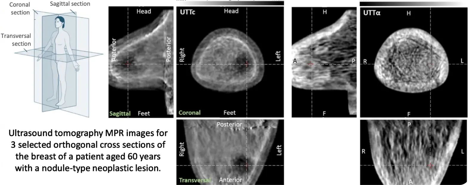
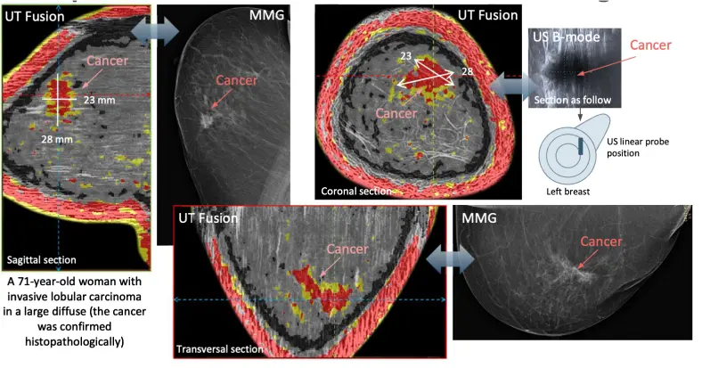
- The images show a neoplastic lesion close to the nipple and near the edge of the breast. There is no significant increase in ultrasound attenuation in the area of the neoplastic lesion, which may indicate that the lump is benign. This lesion was detected automatically in the UT Fusion image (colored tumor areas - yellow with benignity estimation, red with malignancy estimation). Comparative MMG and conventional US imaging confirmed the presence of a similar-sized nodule at the same location with a malignancy estimate of BI-RADS 4a (2 - 10% malignancy). After biopsy, histopathological examination determined that it was a benign leafy FA tumor (fibroadenoma).
Phased array subaperture
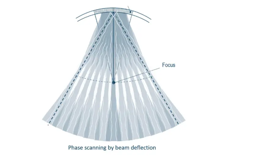
Ultrasound reflection imaging of the scanner uses 32 sectors of an ultrasonic ring array (sub-apertures consisting of each with 32 elementary transducers) as 32 conventional linear B-mode phased arrays. The ultrasound beam is deflected over an angular range of ±30°, with refocusing along an arc so that the focus maintains a distance of about 130 mm. The resulting imaging range for each sub-aperture is approximately 200 mm.
Full angle spatial compound imaging (FASCI) method
The ultrasound reflection imaging mode requires scanning with the activation of 32-transducer sub-apertures sequentially with beam forming and beam steering. By acquiring 32 B-mode images of sectors of the coronal breast cross-section all around, it is possible to reconstruct a two-dimensional image of the entire cross-section by assembling the images with overlap using full angle spatial compound imaging (FASCI) method.
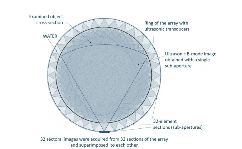
- Large areas of overlap between sectoral images enable pixel averaging, which significantly reduces noise, speckles, artifacts, and increases imaging resolution. A low pulse resonant frequency (2MHz) provides the required imaging long range. The scanning and data acquisition process is switched from transmission mode to reflection mode for each measured coronal breast section to avoid discrepancies due to displacement of breast tissue associated with heartbeat and patient movement.
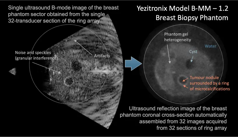
Ultrasound reflection images
- The US FASCI is used in Fusion imaging as a background superimposed on it with appropriate transparency. Transmission images of the distribution of local ultrasound velocity values in breast tissue (UTTc) allow a quantitative assessment of its elastic properties related to density, which is very important because tumor nodules are harder and more elastic compared to surrounding tissue. UTT images of the distribution of local ultrasound attenuation values (UTTα) can be used to refine and increase the likelihood of an accurate diagnosis of cancerous lesions. FASCI images of any breast cross section allow diagnostic reference to the well-known and widely used method of conventional breast ultrasound in medical diagnosis..
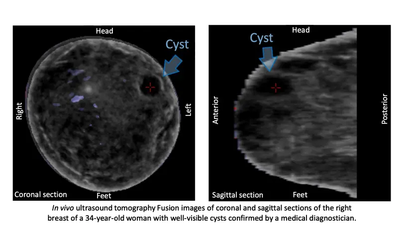
Ultrasound fusion MPR images
- The ultrasound tomography scanner is equipped with a special algorithm, allowing the appropriate fusion of 3 acquired tomographic images (UTTc + UTTα + US FASCI) for any section of the breast. The two transmission images (UTTc and UTTα) are fused with the critical thresholds of ultrasound velocity and attenuation for tumors (taking into account the age of the patient and the individual breast density) and differentiated by colors for fat (black), glandular tissue (blue), benign tumor tissue (yellow if only the ultrasound velocity threshold is exceeded), and malignant tumor tissue (red if both ultrasound velocity and attenuation thresholds are exceeded). An appropriately processed FASCI ultrasound image is used as the background for appropriately pre-assembled transmission images.
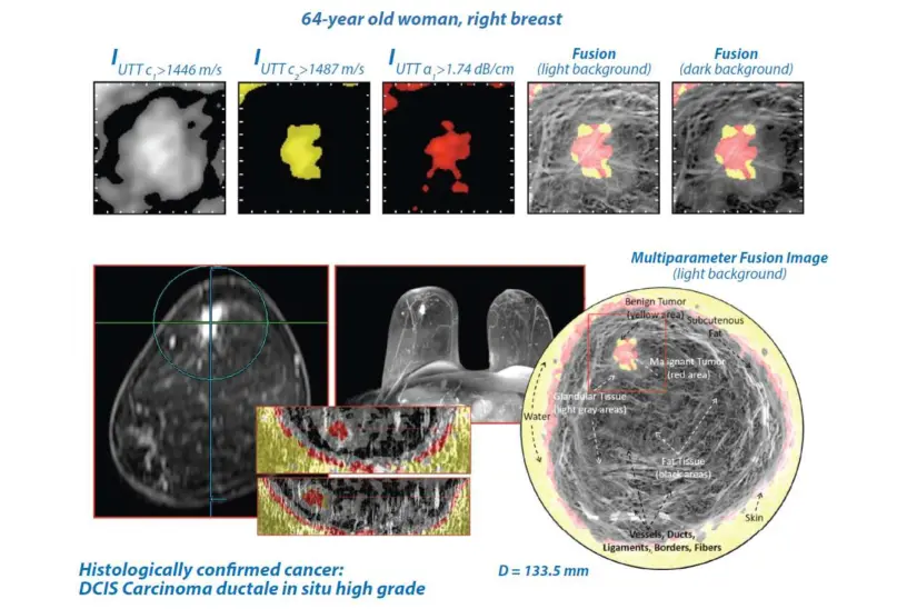
About image resolution
- The longitudinal (axial) and transverse resolution in transmission images (UTT) is limited to a value of 0.4 mm, and the resolution in elevation is limited to about 5 mm. The relatively low vertical resolution (5 mm) is not a disadvantage, as it allows us to increase the shift step of the ring array to 1 mm, thus reducing the time required to scan the entire breast. Pathological changes several millimeters above and below the scanned coronal breast section will still be detected.
- Of decisive importance here is the contrast resolution of the image, which is a determinant of spatial resolution. It depends on the variation of ultrasound velocity and attenuation in the structures of the breast tissue being imaged. Calculations show that using the DRAMIŃSKI S.A. ultrasound tomography scanner, in the case of non-noised ultrasound velocity measurements, it is possible to detect in UTTc images heterogeneities of coronal breast sections < 5 mm in size, differing in ultrasound velocity by at least 1 m/s from the surrounding tissue. The size of detectable inhomogeneities decreases linearly as this velocity difference increases and is limited to 0.5 mm at a velocity difference of 10 m/s due to the limitation of spatial resolution. In UTTα images of coronal breast sections, in the case of non-noised ultrasound attenuation measurements, inhomogeneities with sizes < 2.5 mm that differ in attenuation by at least 10 dB/m from the surrounding tissue can be detected. These sizes decrease linearly with increasing attenuation difference and are limited to values of 0.5 mm at an attenuation difference of 50 dB/m.
-
Opieliński K.J., Pruchnicki P., Szymanowski P., Szepieniec W.K., Szweda H., Świś E., Jóźwik M.,Tenderenda M., Bułkowski M.: Multimodal ultrasound computer-assisted tomography: An approach to the recognition of breast lesions, Computerized Medical Imaging and Graphics, 65, 102-114, 2018. Read more
-
Milewski T., Michalak M., Wiktorowicz A., Opieliński K., Pruchnicki P., Bułkowski M., GieleckiJ., Jóźwik M.: Hybrid Ultrasound Tomography Scanner - a Novel Instrument Designed to Examine Breast as a Breast Cancer Screening Method, Biomedical Journal of Scientific & Technical Research, 14(4), 1-5, 2019. Read more
Contact Us
Phone
+48 89 675 26 11Address
Wiktora Steffena 21
11-036 Sząbruk, Poland
